 the Knee Bone's Connected to the Ankle Bone - Regenexx Blog
AdvertisingRelation between knee and ankle degeneration in a donor population of organs volume 8, article number: 48 (2010) Volume 89328 Accesses25 Citations0 AltmetricAbstractBackgroundOsteoartritis (OA) is a progressive degenerative condition of the synovial joints in response to internal and external factors. The relationship of OA in a union of one limb to another joint within the same limb, or between limbs, has been a topic of interest in reference to the etiology and/or progression of the disease. MethodsThe prevalence of joint lesions of cartilage and osteophytes, characteristic of OA, was evaluated by visual inspection and classification in 1060 adult pairs of knee/tali of 545 cataveric joint donors. ResultsJoint degeneration increased faster with age for knee joints, and significantly more knee joints showed a more severe degeneration than ankle joints since as early as the third decade. Women showed more severe degeneration of the knee than men. The degeneration of the severe ankle did not exist in the absence of severe degeneration of the knee. The effect of the weight on the joint degeneration was specific through which the weight had a significantly greater effect on the knee. The degrees of the ankle did not coincide ever more in a donor, as the degree of degeneration on the left or right knee increased. ConclusionsThe gender and body type have a greater effect on the joint integrity of the knee compared to the ankle, suggesting that the knees are more prone to the internal causative effects of degeneration. We hypothesize that the greatest variability in joint health between joints within an individual as the disease progresses from normal to early degeneration signals can be a result of kinetics of unjusted members, which in turn could lead to the progression of joint diseases. BackgroundOsteoarthritis (OA) is a generally progressive condition that involves anabolic and catabolic mechanisms within the joint cartilage and the bone of the synovial joints in response to internal and external factors. Among these factors are the age [, ] genetic [,], joint alignment/small [-], joint injury [], female gender [] and obesity [, ]. On the other hand, exercise and local muscle strengthening can inhibit their progression [, ] by strengthening the local environment and thus reducing instability in the joint. But because there is no cure for the OA and because its etiology is not fully understood, research continues to dilute the mechanisms that can contribute to its initiation and progression. The alignment of the tomb and the relationship between biomechanics, as well as the incidence/prevalence of the OA in a union of an extremity in relation to another articulation within the same limb has been a topic of re-emerging interest. Recently, a cross-sectional study [] found multiple kinematic and kinetic differences in the hip, knee and ankle joints in those individuals with severe OA knee. In addition, using mechanical axis x-rays, Tallroth et al. [] found that the higher the inclination ( relative crayon to distal tibia and distal fibula) in the ankle, the more degenerative the changes. Previously, on a sample of 50 knee and ankle donors, it was shown that donors with ankle degeneration also showed degenerative changes within the knee at an equal or higher level []. Data suggest that factors such as altered mechanics as a result of extremity alignment could contribute to degeneration within a whole limb. Furthermore, although genetic factors might be involved in the global joint degeneration within an individual, mechanical factors within a member probably influence the joint health of the counter-lateral member. The aim of this study was to expand our previous database [] and correlate knee and ankle stitches OA in an effort to further clarify the relationship between degenerative joint disease within a member and between members of an individual. Hypothesis that AO in a joint is associated with a higher prevalence of AO in another kinematically related joint, with this relationship increasing with the severity of AO. MethodsA thousand sixty pairs of adults on the knee/tali of 545 joint donors were obtained through collaboration with the Donors of Hope Network and the Tissue Donor Network. This included two knees and two ankles of each catavric donor with the exception of 30 donors for which only one lower limb was available because the other was present but not available to our laboratory. The joints were collected between June 1995 and April 2009 in accordance with the Gift of Hope policies and with the approval of the Institutional Review Board of Rush University. Exclusion criteria included prior amputation of a limb, joint replacement in a lower limb, hepatitis history, or postmortem positive blood test for hepatitis, HIV, or any other communicable disease. The distal portion of the thybia (proximal component of the ankle) was not available; therefore, the talus represents the joint of the ankle in this study. Although there are no medical stories of donors, age, gender and cause of death are provided. Donors were classified as light, normal or obese based on the subjective visual assessment of the joint harvester. The joints were opened within 24 hours of the donor's death and were examined for the interruption of the articular cartilage in a modified Collins scale [] where grade 0 is normal smooth cartilage, grade 1 is superficial fibrillation, grade 2 is superficial fissure or ulceration with possible osteophytes, grade 3 is 30% or less of the surface of eroded cartilage to the most carotid bone Figure 1 Representative Tali. Examples of joint degeneration degrees: (a) grade 0, (b) grade 1, (c) grade 2, (d) grade 3, (e) grade 4.Representative account (a) (b) (c) (d) (e) The following statistical analysis was carried out for non-parametric data. Spearman's range correlation was carried out to determine the correlation between age and joint degeneration for each joint individually. The range test signed by Wilcoxon was used to determine the relationship between the degree of the left and the right for the ankle and knee joints separately. To determine the effects of tissue type (ancle vs. knee) and sex in age-dependent degeneration, a survival analysis was performed. The analysis determined the incidence (such as survival rate) of the selected grades (set to 1 to 4) older (i.e., "survival curve"), stratified by tissue type. The average age of survival, the age at which half of the samples were degenerated (i.e., it became the selected grade), was also determined. Survival curves were compared to the Kaplan Meier analysis and the Mantel statistics []. In addition, the effect of sex on survival curves was determined separately for each type of tissue. Statistical importance was taken in P ResultsDonors ranged from 19 to 98 years of age at an average age of 60 years. The distribution per decade is shown in Figure . There were 287 men (53%) and 258 (47%) women. This profile reflects the donor population of the Tissue Donor and Donor Network. Figure 2 Distribution of knee/tali donors per decade of life. Average age = 60 years. Distribution of knee/tali donors per decadeIn the current study population, joint degeneration was first observed during the third decade of life, starting with a 26-year-old male with both knees showing fibrilled cartilage (grade 1). The first signs of degeneration in the ankles were in a 28-year-old male whose knees showed grade 2 and the ankles showed degeneration of grade 1. The oldest donor, a 96-year-old woman had knees with grade 4 degeneration and ankles with grade 2 degeneration. The percentage of donors in each decade showing each degree of degeneration for knees and ankles is shown in Figures and , respectively. We found that 17% of the individuals had both the normal knee (grade 0) and the ankle cartilage. Degeneration increased in ankles over the tenth decade. Spearman's range correlation for the association between age and degeneration level in the left and right joints of the ankle and the knee was statistically significant (P = 0.0001 for both joints). Degeneration increased in the knees during the ninth decade, with a slight decline in the tenth decade, but it should be noted that there were only six donors in the tenth decade. Degeneration increased faster with age for knee joints, and significantly more knee joints showed a more severe degeneration than ankle joints since as early as the third decade. A minority (4 per cent) of knee joints showed completely normal cartilage from the sixth decade to the top, and no knee joint shows normal cartilage for the eighth decade. For the ankle, however, approximately 50% of the joints had completely normal cartilage over the sixth decade. Erosion of 30% or less of the cartilage surface (grade 3) began as early as the fourth decade for the knees and the fifth decade for the ankles. The diffuse erosion of cartilage (grade 4) began as early as the fourth decade for the knees, but it was only loosened in three donor ankles and not until the eighth decade. Over the eighth decade, the majority (63%) of the ankles continued to show normal cartilage or fibrillation, while at this time, most of the knees showed moderate to severe degeneration. Figure 3 (a) and (b) Distribution of grades per decade for (a) knee joints and (b) ankle. In the third decade, the severity of the knee notes increased compared to the ankle. In the eighth decade, there was no normal knee in appearance, while in the tenth decade there was some normal tali.(a) and (b) Distribution of grades per decade to (a) knee joints and (b) ankles The knee and ankle cartilage scores, separately, for the left and right sides are shown in Figure . There were no significant differences between sides (P Ø 0.05). Most individuals had a more severe OA knee than the OA ankle (60.8% on the left side and 60.5% on the right side). Less individuals had equal AO severity on the knee and ankle (30.1% on the left side and 36.9% on the right side). Rarely the ankle showed AA more severe than the knee (1.1% on the left side and 2.6% on the right side). Figure 4 Distribution of grades for left and right knees and ankles separately. Grade distribution for left and right knees and ankles separatelyResults from Wilcoxon's signed range test revealed that the only joint relationships that showed no significant differences were left vs.right ankles and left right knees vs. (P = 0.1705 and 0.0845, respectively). The figure shows cartilage scores by gender, where it can be seen that slightly less knees and female ankles were normal (grade 0) than male knees and ankles (P = 0.05). Females also showed a more severe degeneration of the knee (grades 3 and 4) than males (P = 0.03). Figure 5 Distribution of grades for male and female knees and ankles separately. Distribution of the categories of male and female knees and ankles separately The effect of the weight on the joint degeneration was significant (Figure ). The knees of the obese individuals showed more severe (grades 3 and 4) degeneration than the knees of normal or lightweight donors (P 0.05 for grades 3 and 4). Figure 6 Distribution of the knee and ankle grades separated according to the relative body type (as visually evaluated). The knees of obese donors showed a more severe degeneration (grades 3 and 4) than the joints of light or normal donors. Distribution of knee and ankle grades separated according to relative body type (according to visual evaluation) The relationship between knee and ankle degeneration within individual donors can be resolved in several ways. A review of which joint degeneration shows more serious than its counterpart within an individual's limb shows that the majority (61%) of donors had knees more severely degenerated than ankles on the left and right sides. Less (38.1% [on the left], 36.9% [on the right]) had knees and ankles of the same degree of degeneration, and only 1.1% (on the left) and 2.6% (on the right) had ankles that were more degenerated than the knees. This was true for the left and right extremities. 90% of donors with normal knees (grade 0) also had normal ankles, while 38% of donors with normal ankles also had normal knees. For a deep look at the most severe degeneration, the Table shows the percentage of ankle joints in each degree of cartilage when the ipsilateral/contralateral knee joint showed degeneration of grade 4, and vice versa for the opposite joint. From this table it is evident that people with severely degenerated knees may have normal ankle cartilage; this was the case as often as their coated or insured ankle cartilage. However, many less ankles showed the same severity of degeneration as the ipsilateral or contralateral knee, although this condition did exist. Although there were only three ankles showing degeneration grade 4, they were associated with degenerated knees. Table 1 Percentage of ankle joints at each cartilage grade when the knee joint showed degeneration of grade 4 (the most severe degeneration)Looking at the relationship between the joint degeneration as the lowest/highest joint of the limb changed in health condition, Table shows the distribution of the paired and unmatched ankle grades within a donor with different levels of degeneration. The ankles within a donor did not coincide more and more (i.e. they did not have the same degree) as the degree of degeneration on the left knee increased through grade 3, with a little decrease in grade 4 (P Table 2 Distribution of the matching and unmatched ankles in association with the different levels of joint degeneration within the knee of the same donor If both knees showed grades of 3, 63,8%). If both knees showed grades of 4, 81.8% (18 of 22) of these ankles had the same degree. There were 11 donors (23.4 per cent of the grade 3 donors in which both knees were grade 3 and both ankles were grade 3 0. There were five donors (22.7 per cent of grade 4) where both knees were grade 4 and both ankles were grade 0. Survival analysis to compare ankles and knees (Figure) suggested a growing incidence of degeneration with aging both for knees and ankles, but markedly different survival curves for the ankle compared to the knee (P Figure 7) Survival curves of the ankle (red) and the knee (blue), which reflect the likelihood that the samples reach grades (a) 1, (b) 2, c) 3 or d) 4. There is a significant difference between the expected survival of the knees and ankles (P = 0.001). Survival curves of the ankle (red) and the knee (blue), reflecting the likelihood that the samples reach grades (a) 1, (b) 2, (c) 3 or (d) 4 Survival analysis to evaluate sexual differences (fig.) suggests significant differences between male and female knee survival curves when grade 3 (fig.) or 4 was defined as degenerating. However, there was a trend (P = 0.09) of relatively retarded degeneration (degree 2) in women's ankles between 80 and 95 years of age (Figure). Figure 8 Survival curves of male (red) and female (blue) samples in (a) knees reaching grade 3 and (b) ankles reaching grade 2. There were significant differences between the curves of male and female knees when grade 3 (figure 8a) or 4 was defined as degenerate (for each P survival curves of male (red) and feminine (b) samples in the knees reaching grade 3 and (b) ankles reaching grade 2DiscussionOA is a condition based on both degenerative changes in the carti. In this study, because the history of the pain of individuals within our donor population was not available, we do not use the term osteoarthritis, but the joint degeneration []. This is important because it is well known that some individuals with joint pain do not show evidence of radiographic or magnetic resonance of the joint disease, while other individuals without joint pain show evidence of images of pathological joint changes normally associated with OA [, ]. One force of this study, however, is that we had the advantage of real visualization of articular cartilage surfaces and osteophytes of cataverous cross section donors, thus giving data on normal and early stages of the disease that cannot be discerned through any current imaging technology. In a very early analysis of our donor population, when only 50 pairs of knee donors or anchors had been harvested, we found that the joint degeneration of the ankle was more frequent in men than in women, it increased with age, and was more often given in both members with the same gravity []. In donors with ankle degeneration, the knee also showed degenerative changes with an equal or higher degree. At that time, we suggest that factors such as altered mechanics could be responsible for degeneration in an extremity and produce changes in the contralateral extremity. The present study on 545 knee and knee donors with an average age of 60 years reaffirms our previous results. For the joint of the knee, females showed greater degeneration than males, with less normal joints and more joints that show partial and extreme erosion of the joint surface. This coincides with the well-known highest prevalence of AO in women compared to men [, ]. This difference can be due to one or more of several known gender differences involving knee joint anatomy, kinematics and/or physiology [–]. A difference we found compared to a previous study [] was that here donors had a slightly less normal ankle cartilage and a little more fibrillation than males. However, this was not extrapolated to higher degrees of degeneration, where male ankles showed a slightly earlier fissure (grade 2) than female ankles. The effect of the weight on the joint degeneration was specific through which the weight had a significantly greater effect on the knee than on the ankle. Most of the knees of obese donors showed degeneration of at least grade 2 (fissuring) or greater, and nearly 50% showed cartilage erosion to the subcondral bone. In the ankle, although light donors showed little fissure and no erosion, the levels of fissure and erosion were not different between individuals with normal and obese weight. We found that approximately 20% of donors in which both knees showed advanced degeneration (grades 3 or 4) had ankles that seemed perfectly normal; the reverse never happened. This reinforces the idea that knee degeneration will probably have a greater influence on ankle health than the reverse situation. The fact that the knees can be bilaterally severely pathological in the structure in the absence of visible ankle pathology attests to the structural stability of the ankle as a hinge joint with less mechanical freedom compared to the knee. It seems that, at least in some individuals, the structure and function of aberrant knees inevitably do not lead to changes in the function of the limb so severe that they affect the ankle. At a purely speculative level, however, it is likely that this protection will not be observed on the hip, as the hip is much more prone to the OA than the ankle, and the coexistence of the hip and the OA knee is well documented [, ]. Survival analysis suggests that even mild degeneration (grade 1) occurs more slowly in the ankle than in the knee, and severe degeneration (grades 3 and 4) rarely occurs in the ankle. Furthermore, the effect of sex on joint degeneration was specific and depended on the severity of degeneration. On the knee, mild to moderate degeneration (grades 1 and 2) occurred similarly in both sexes; however, severe degeneration (grades 3 and 4) occurred at an early age for women. The trend was reversed in the ankle. In addition, we explored the data a little different to further clarify the relationship between the two joints within a limb. An interesting relationship occurred when seeing how degeneration on the knee was related to pairing the grades of the ankle within an individual. The grades of the ankle did not more and more coincide with a donor, since the degree of joint degeneration on the left knee increased through grade 3 (partial erosion of the joint surface), with a decrease in grade 4 (total erosion). There was an increase in the number of grades of ankle unmatched as the right knee degeneration increased through grade 2 (fissuring), at which time the percentage of grades remained basically uneven. This points to increased variability in joint health within the limbs and leads to an imbalance in joint health between the sides as the disease progresses. Combined with the finding that since degeneration on the knee increased thus degeneration on the ankle, an interesting consideration appears. 90% of donors with normal knees (grade 0) also had normal ankles, while 38% of donors with normal ankles also had normal knees. However, once signs of degeneration of the knees occur, even in the early stages (i.e., fibrillation), the ankles of a pair begin to be discordant in their appearance with respect to others. We interpret this as suggesting that any mechanism is occurring on the knee to cause early degeneration, the same mechanism probably occurs on the ankle, but at a lower level. This can be the result of mechanical alterations in the knee or through an independent process. It is likely, however, that both are related as suggested in studies that have attempted to dilute the relationship between the knee and the OA ankle. Studies in patients have shown that the alignment of rubber-ankle contributes to the distribution of the load through a joint surface. In fact, both varo and valgus malignment of the knee increase the risk of progression of OA medial and lateral, respectively [, ]. The rod knee increases strength through the middle knee compartment, while the side compartment has increased the strength in the valgus knee []. In both conditions, the mechanical alignment of the extremity is changed from the neutral axis, thus establishing alignment problems along the limb and perhaps the whole body. In a retrospective study of mechanical axle x-rays just before the total knee arthroplasty, it was found that the OA ankle and the slope in the ankle were not uncommon []. Moreover, the higher the inclination in the ankle, the more degenerative the changes in the joint []. When the mechanical axis was corrected on the knee at the time of surgery, the ankle inclination was also significantly changed. This work relates well to one of our previous studies in which we find that the trabecular angle within the dome of the drill is associated with the level of joint degeneration []. The dome of the human talus receives compressive forces that have crossed the leg. Thus, according to the Wolff Law, the body of the talus has predominated vertically lined trabecula that functions superior to inferior. Through the rapid Fourier transformation analysis, it was found that, as the trabecular angle deviates from a perpendicular alignment, the largest was the cartilage changes on the joint surface, particularly on the medial and lateral borders. Hypothesis that these results can be a reflection of the alignment and/or biomechanical in the joint []. Therefore, taking the ideas of these last two studies together, it is possible that a bad knee affects the alignment of the entire kinetic chain, setting the stage for potential pathology anywhere along that chain. Another relationship that would have been interesting to examine is how medial vs. the OA side knee is related to the medial ankle vs. lateral OA. Unfortunately, because we did not have information about the topographic location of cartilage changes, we cannot make any statements about it. However, in a previous cavric study, we found that more knee and ankle joints showed greater degeneration in the medial than in the side aspect []. In another study, on the difference between the standing center of pressure patterns between subjects with and without OA, we find that the subjects with medial compartment OA showed a more laterally placed foot pressure pattern with normal walking compared to non-OA control subjects []. This is achieved by changing the axis of the ankle joint relative to the leg and exerting greater pressure on the mid-size aspect of the ankle. Therefore, at least of these results, it could be expected that the OA mid ankle could be found in relation to the OA knee. However, further studies should be undertaken to determine this determination. The limitations of this study include the lack of information on the history of joint damage and the lack of information on the level of mobility or the use of walking aids. Each of these issues has the potential to introduce variability in data that may not be accounted for. For example, if a subject suffered an undocumented traumatic injury in the knee joint, it would not be known if the presence of OA in this joint was due to a trauma or the relationship of this joint with the contralateral knee or ankles. Another limitation is that we had no access to distal warmth. If the joint degeneration in this component is greater than that of the same set, this can lead to the underestimation of the true severity of the ankle pathology. This would surely be the case in at least some specimens, as we have shown earlier in a sample of 100 specimens of 50 corpses that 30% of ankle joints showed greater degeneration in the tibia than in the talus, 21% showed equal levels of degeneration on both sides and 49% showed greater degeneration in the talus []. Another consideration parameter is the way the body type was determined (light, normal, obese). Because we got the joints through the Tissue Donor and Organ Hope Network, we depend on subjective determination after physical examination of the body. We consider the amount of general subcutaneous body fat when making this determination, and although not totally scientific, we believe that this method provides good relative information within the study sample. Conclusions For our knowledge, this joint study of Catalan donors is the largest study of its type of pathology of knees and ankles. The joint of the knee showed signs of degeneration significantly greater than the joint of the ankle and showed a gender preference for which women had a more severe knee degeneration. Obesity increased the severity of joint lesions on the knee but had a much less profound effect on the ankle. A new important finding of the study was that the grades of ankle did not coincide more and more in a donor, as the degree of joint degeneration on the left or right knee increased. This is, in essence, an imbalance in the joint integrity of the parties and points to greater variability in joint health within the limbs as the disease progresses. The possibility that this will lead to the misleading of the limbs, especially once the cartilage erosion has occurred in at least one knee compartment, is realistic. In turn, the malignment of the limbs is very associated with the progression of joint diseases as shown in other studies. ReferencesGensburger D, Arlot M, Sornay-Rendu E, Roux JP, Delmas P: Radiological assessment of changes in the joint knee space related to age in women: a 4-year longitudinal study. Rheum arthritis. 2009, 15: 336-343. 10.1002/art.24342. Loeser RF: aging and osteoarthritis: the role of condrocytic senescence and aging changes in the cartilage matrix. Cartilage osteoarthritis. 2009, 17: 971-979. 10.1016/j.joca.2009.03.002. MacGregor AJ, Li Q, Spector TD, Williams FM: Genetic influence on radio osteoarthritis is a specific site in the hand, hip and knee. 2009, Rheumtology (Oxford), 48: 277-280. Issa SN, Dunlop D, Chang A, Song J, Prasad PV, Guermazi A, Peterfy C, Cahue S, Marshall M, Kapoor D, Hayes K, Sharma L: Complete tomb X-ray and knee-shot evaluations of varus-valgus alignment and its relationship with the characteristics of osteoarthritis disease by magnetic resonance. Rheum arthritis. 2008, 57: 398-406. 10.1002/art.22618. Kalichman L, Zhu Zhang Y, Niu J, Gale D, Felson DT, Hunter D: The association between the alignment of the patella and the pain and function of the knee: a study of MRI in people with symptomatic knee osteoarthritis. Cartilage osteoarthritis. 2007, 15: 1235-1240. 10.1016/j.joca.2007.04.014. Hunter DJ, Niu J, Felson DT, Harvey WF, Gross KD, McCrea P, Aliabadi P, Sack B, Zhang Y: Knee alignment does not predict incident osteoarthritis: the study of Framingham osteoarthritis. Rheum arthritis. 2007, 56: 1212-1218. 10.1002/art.22508. Hunter DJ, Sharma L, Skaife T: Knee alignment and osteoarthritis. J Bone Joint Surg (Am). 2009, 91 (S1): 85-89. 10.2106/JBJS.H.01409. Tanamas S, Hanna FS, Cicuttini FM, Wluka AE, Berry P, Urquhart DM: The malignment of the knees increases the risk of development and progression of osteoarthritis of the knees: a systemic review. Rheum arthritis. 2009, 61: 459-467. 10.1002/art.24336. Englund M, Guermazi A, Roemer FW, Aliabadi P, Yang M, Lewis CE, Torner J, Nevitt MC, Sack B, Felson DT: Mensical tear on the knees without surgery and development of radiographic osteoarthritis between middle-aged people and elderly people: The multicenter study of osteoarthritis. Rheum arthritis. 2009, 60: 831-839. 10.1002/art.24383. Hanna FS, Teichtahl AJ, Wluka AE, Wang Y, Urquhart DM, English DR, Giles GG, Cicuttini FM: Women have increased rates of cartilage loss and progression of cartilage defects on the knee than men: a gender study of adults without clinical knee osteoarthritis. Menopause. 2009, 16: 624-625. 10.1097/gme.0b013e318198e30e. Grotle M, Hagen KB, Natvig B, Dahl FA, Kvien TK: Obesity and osteoarthritis in the knee, hip and/or hand: an epidemiological study in the general population with 10 years of follow-up. BMC Musculoskelet Disord. 2008, 9: 1-5. 10.1186/1471-2474-9-132. Claessen H, Arndt V, Drath C, Brenner H: Overweight, obesity and risk of labor disability: a cohort study of construction workers in Germany. Occup Environ Med. 2009, 66: 402-409. 10.1136/oem.2008.042440. Hunter DJ, Eckstein F: Exercise and osteoarthritis. J Anat. 2009, 214: 197-207. 10.1111/j.1469-7580.2008.01013.x. Amin S, Baker K, Niu J, Clancy M, Goggins J, Guermazi A, Grigoryan M, Hunter DJ, Felson DT: Quadriceps strength and risk of loss of cartilage and progression of symptoms in knee osteoarthritis. Rheum arthritis. 2009, 60: 189-198. 10.1002/art.24182. Astephen JL, Deluzio KJ, Caldwell GE, Dunbar MJ: Biomechanical changes in the knee of the hip and ankle joints during the gait are associated with the severity of the osteoarthritis of the knee. J Orthop Res. 2008, 26: 332-341. 10.1002/jor.20496. Tallroth K, Harilainen A, Herttula L, Sayed R: The osteoarthritis of the ankle is associated with osteoarthritis of the knees. Conclusions based on mechanical axis X-rays. Arch Orthop Trauma Surg. 2008, 128: 555-560. 10.1007/s00402-007-0502-9. Koepp H, Eger W, Muehleman C, Valdellon A, Buckwalter JA, Kuettner KE, Cole AA: Prevalence of joint cartilage in the ankle and knee joints of human organ donors. J Orthop Sci. 1999, 4: 407-412. 10.1007/s007760050123. Muehleman Bareither D, Huch K, Cole AA, Kuettner KE: Prevalence of degenerative morphological changes in the joints of the lower extremity. Cartilage osteoarthritis. 1997, 5: 23-37. 10.1016/S1063-4584(97)80029-5. Mantel N: Evaluation of survival data and two new rank order statistics that arise in its consideration. Cancer Chemother Rep. 1966, 50 (3): 163-170. Bedson J, Croft PR: The discordance between clinical and radiographic knee osteoarthritis: a systematic search and a summary of literature. BMC Musculoskelet Disord. 2008, 9: 116-10.1186/1471-2474-9-116. Dieppe P, Judge A, Williams S, Ikwueke I, Guenther KP, Floeren M, Huber J, Ingvarsson T, Learmonth I, Lohmander LS, Nilsdotter A, Puhl W, Rowley D, Thiler R, Dreinhoefer K: Variations in the preoperative state of patients arriving at the primary hip of osteoart. 2009, 10: 10-19. 10.1186/1471-2474-10-19. Peyron JG, Altman RD: The epidemiology of osteoarthritis. In Osteoarthritis: Diagnosis and Medical/Burgic Management. Edited by: Moskowitz RW, Howell DS, Goldberg VM, et al. 1992, Philadelphia: W.B. Saunders, 15-37. 2 Srikanth VK, Fryer JL, Zhai G, Winzenbert TM, Hosmer D, Jones G: A meta-analysis of the prevalence of sexual differences, incidence and severity of osteoarthritis. Cartilage osteoarthritis. 2005, 13: 769-81. 10.1016/j.joca.2005.04.014. Cushnaghan J, Dieppe P: Study of 500 patients with joint osteoarthritis of members. I. Age analysis, sex and distribution of symptomatic joint sites. Ann Rheum Dis. 1991, 50: 8-13. 10.1136/ard.50.1.8. Wojtys EM, Ashton-Miller JA, Huston LJ: A gender-related difference in the contribution of the musculature of the knee to the stiffness of the sagittal clairy in subjects with laxity of the similar knee. J Bone Joint Surg (Am). 2002, 84 (A1): 10-16. Csintalan RP, Schulz MM, Woo J, McMahon PJ, Lee TO: Gender differences in the biomechanical articulate paellofemoral. Clin Orthop Relat Res. 2002, 402: 260-269. 10.1097/00003086-200209000-00026. Hsu WH, Fisk JA, Yamamoto Y, Debski RE, Woo SL: Differences in the torsional joint stiffness of the knee among the genres: a human body study. Am J Sports Med. 2006, 34: 765-770. 10.1177/0363546505282623. Dıraçoğlu D, Alptekin K, Teksöz B, Yağcı I, Ozçakar L, Aksoy C: Injecting intra-articular hyaluronic acid in patients with hip and knee osteoarthritis: a pilot study. Clin Rheumatol. 2009, 28: 1021-1024. 10.1007/s10067-009-1199-7. Allen KD, Coffman CJ, Golightly YM, Stechuchak KM, Keefe FJ: Daily variations of pain among patients with hand, hip and knee osteoarthritis. Osteoarthritis Cartillage. 2009, 17: 1275-1282. 10.1016/j.joca.2009.03.021. Sharma L, Song J, Felson DT, Cahue S, Shamiyeh E, Dunlop DD: The role of the alignment of the knees in the progression of diseases and the functional deterioration of osteoarthritis of the knees. JAMA. 2001, 286: 188-195. 10.1001/jama.286.2.188. Erratum in: JAMA 2001, 286: 792 Brouwer GM, van Tol AW, Bergink AP, Under JN, Bernsen RM, Reijman M, Pois HA, Bierma-Zeinstra SM: Association between valgus and varus alignment and the development and progression of radiographic osteoarthritis of the knee. Rheum arthritis. 2007, 56: 1204-1211. 10.1002/art.22515. Tetsworth K, Paley D: Malalignment and degenerative arthropathy. Orthop Clin North Am. 1994, 25: 367-377. Schiff A, Li J, Inoue N, Masuda K, Lidtke R, Muehleman C: The trabecular angle of the human story is associated with the degeneration level of cartilage. Interact Neuronal Musculoskelet. 2007, 7: 224-230. Lidtke R, Muehleman C, Block JA: The foot pressure center is related to medial osteoarthritis of the knees. J Am Podiat Med Assoc. 2010, 100: 178-184. Pre-publication history The pre-publication history of this document can be accessed here: Recognition We are grateful to the Illinois Donor Network of Hope and the Tissue Donor Network and to the families of donors. Author's information Affiliates Department of Biochemistry, Rush University Medical Center, Cohn Research Building, 1735 W. Harrison, Chicago, IL, 60612, USACarol Muehleman & Arkady MargulisDepartment of Radiology University of California, San Diego, 11379 Cadence Grove Way, San Diego, CA, 92130, USAWon C BaeDepartment of Orthopaedic Surgery, You can also search this author in You can also search this author in You can also search this author in Correspondence to the correspondent . Additional Information Competing interests The authors declare that they have no competing interests. Contributions by authors CM analyzed the data and wrote the manuscript. AM harvested and qualified the human knees and tali from 2004 to 2009. WB performed the survival analysis, prepared some of the figures and contributed to the writing of the manuscript. KM performed statistical analysis and contributed to the writing of the manuscript. Arkady Margulis, Won C Bae and Koichi Masuda also contributed to this work. Original files presented by the authors for images Below are the links to the original files presented by the authors for images. Rights and permits This article is published under license to BioMed Central Ltd. This is an Open Access article distributed under the terms of the Creative Commons License ( Creative Commons), which allows unrestricted use, distribution and reproduction in any medium, provided that the original work is duly cited. About this article Cite this articleMuehleman, C., Margulis, A., Bae, W.C. et al. Relationship between knee and ankle degeneration in a population of organ donors. BMC Med 8, 48 (2010). https://doi.org/10.1186/1741-7015-8-488, Received: 02 July 2010Accepted: 28 July 2010Published: 28 July 2010DOI: KeywordsWarning BMC Medicine ISSN: 1401-7015 Contact us BMC By using this website, you agree to our , , and politics. We use at the center of preferences. © 2021 BioMed Central Ltd unless otherwise indicated. Part of .
Accessibility Links Search ModesSearch Results2.972 How the human knee works - MITBiomecánica de lo natural, artrtico y replaced human ...
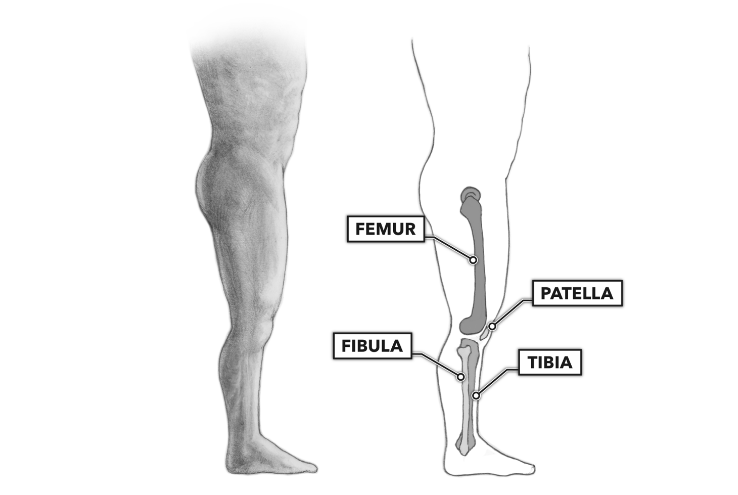
CrossFit | Movement About Joints, Part 6: The Knee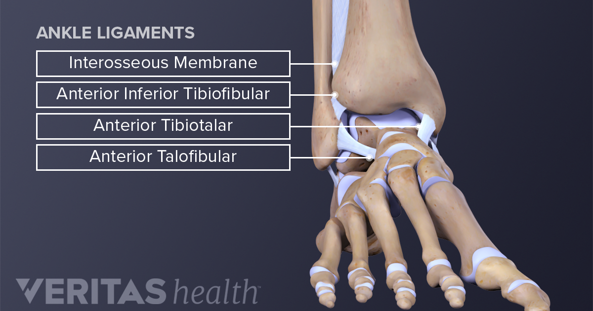
Ankle Anatomy: Muscles and Ligaments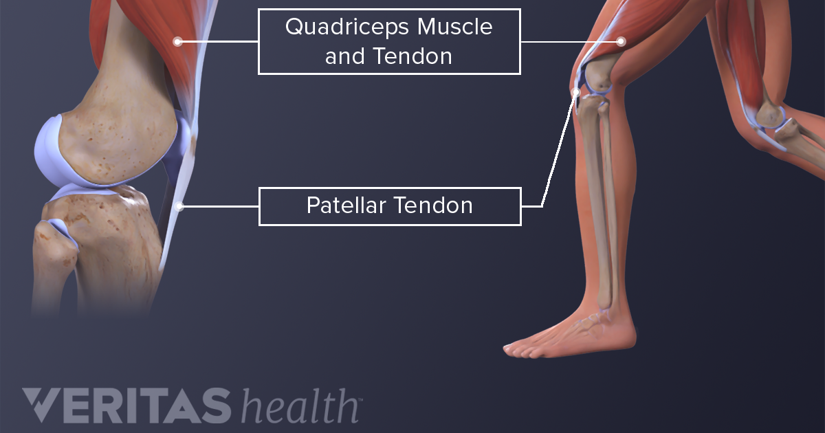
Understanding Jumper's Knee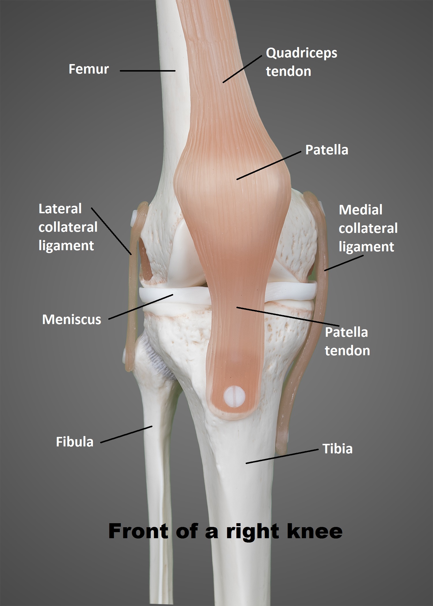
The Knee | UT Health San Antonio
The Foot & Ankle | Global Alliance for Musculoskeletal Health
Knee Hyperextension: Its All In Your Mind! — Ellie Herman Pilates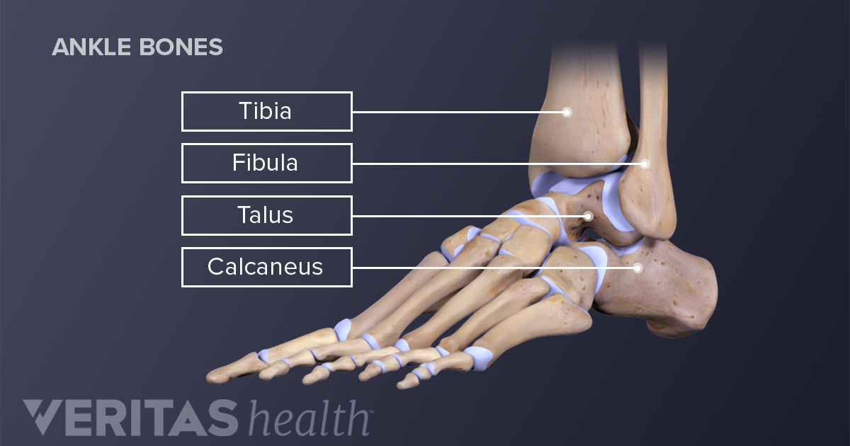
Ankle Joint Anatomy and Osteoarthritis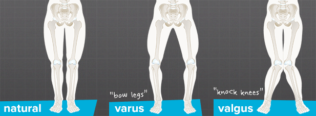
What is the Mechanical Axis of the Knee? - Brainlab.org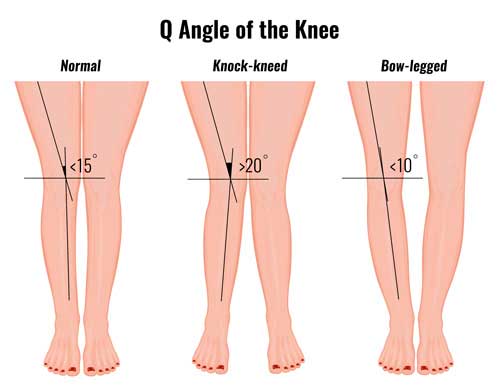
Q Angle & Knee Rehabilitation - Sportsinjuryclinic.net
Decreased Ankle Dorsiflexion is Associated with Dynamic Knee Valgus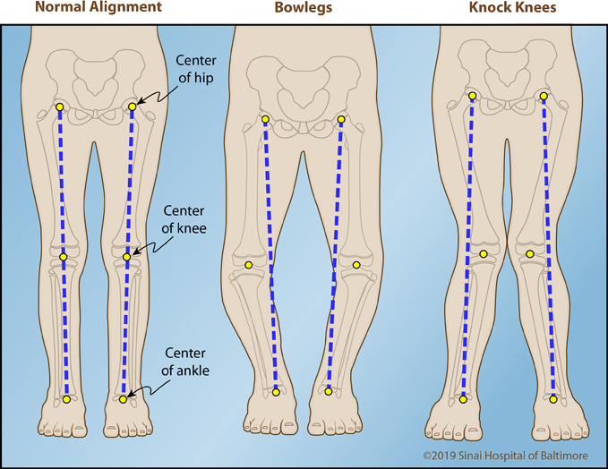
Bowlegs | International Center for Limb Lengthening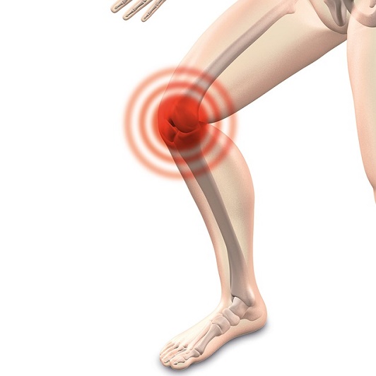
Why Does My Knee Feel Like It Wants to Pop?
Types of Knee Pain: Anterior, Posterior, Medial, & Lateral
Knee | Clinical Gate
Knee - Physiopedia
Decreased Ankle Dorsiflexion is Associated with Dynamic Knee Valgus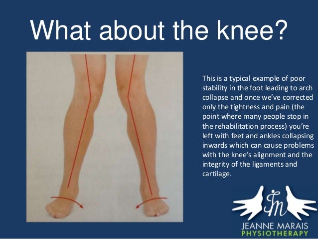
The impact of and rehab for a Bad Ankle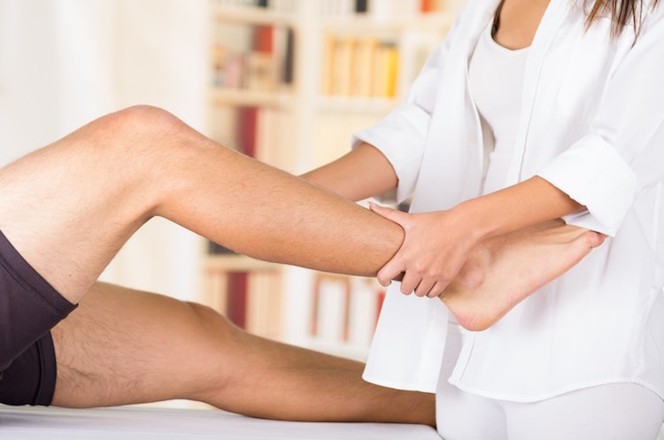
Leg (knee to ankle) - superficial posterior view - MyDr.com.au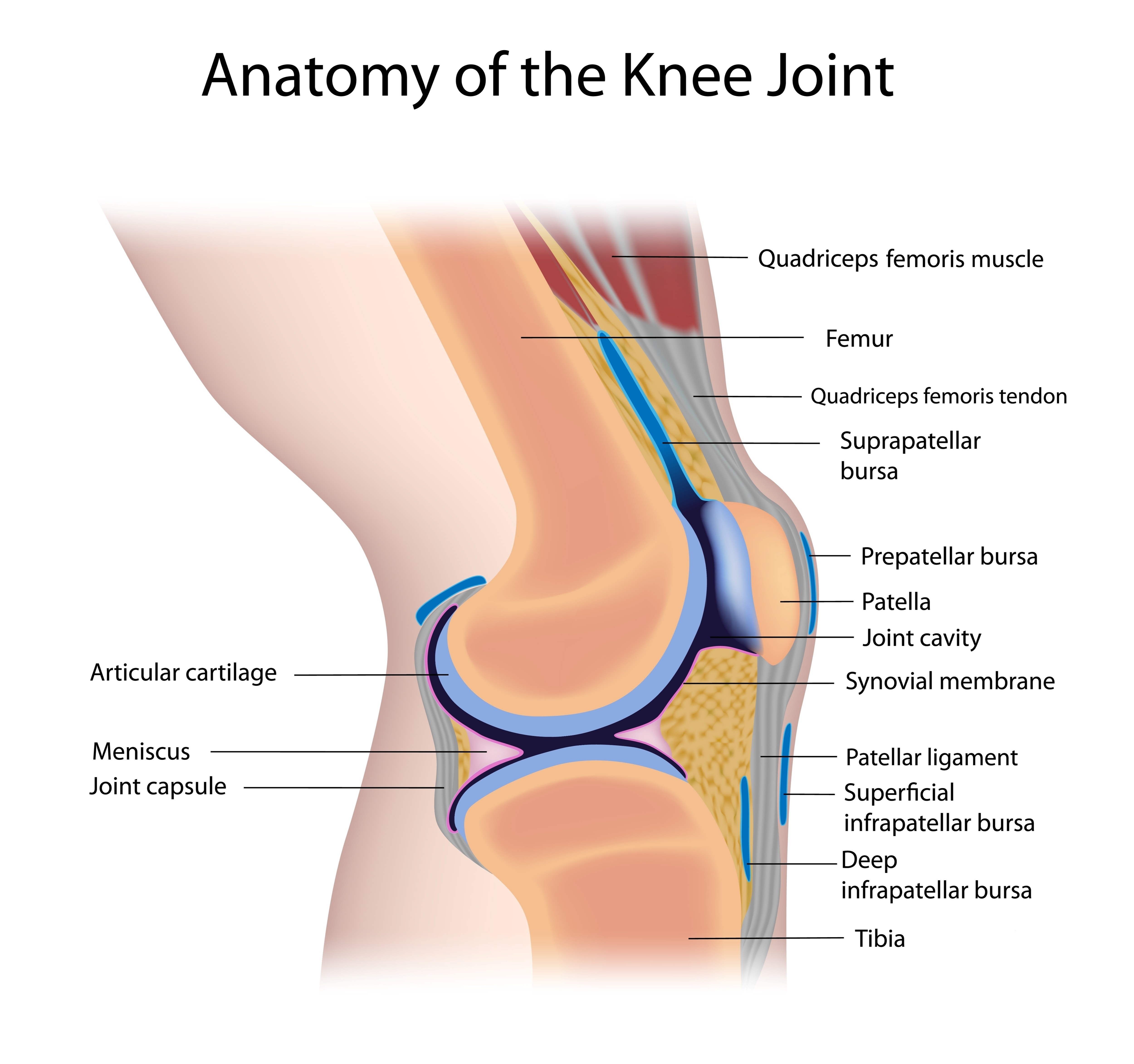
How to Protect Your Knees and Make Them Healthy – SAPNA Pain Management Blog/kneepainwhenrunning-58d2ae2b3df78c5162051537.jpg)
Why Do I Feel Knee Pain When Running?
Can a Sprained Ankle cause Knee Pain?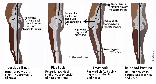
Knee Hyperextension: Its All In Your Mind! — Ellie Herman Pilates
What is a rotationplasty? - YouTube:background_color(FFFFFF):format(jpeg)/images/library/11077/175_176_177_178_179_tibia_extensors_nn.png)
Leg and knee anatomy: Bones, muscles, soft tissues | Kenhub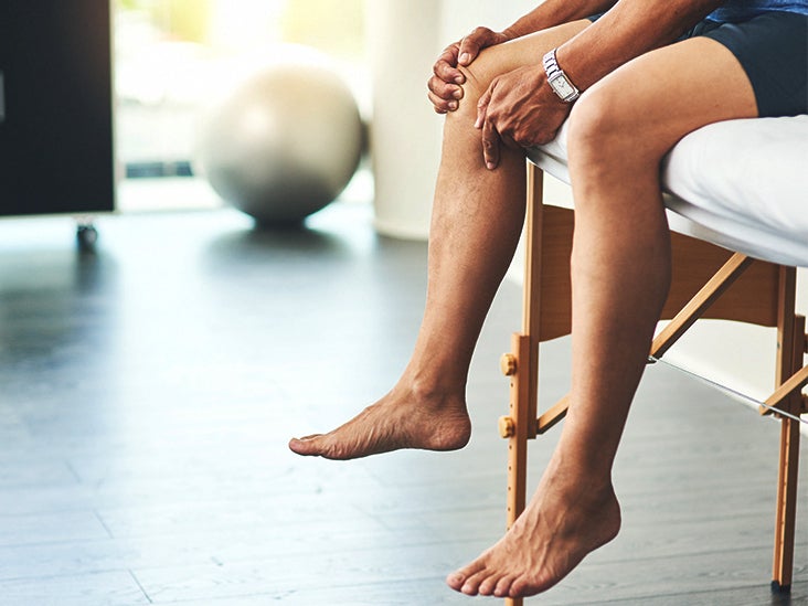
Joint Pain: Causes, Home Remedies, and Complications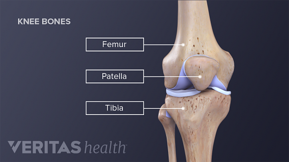
Knee Anatomy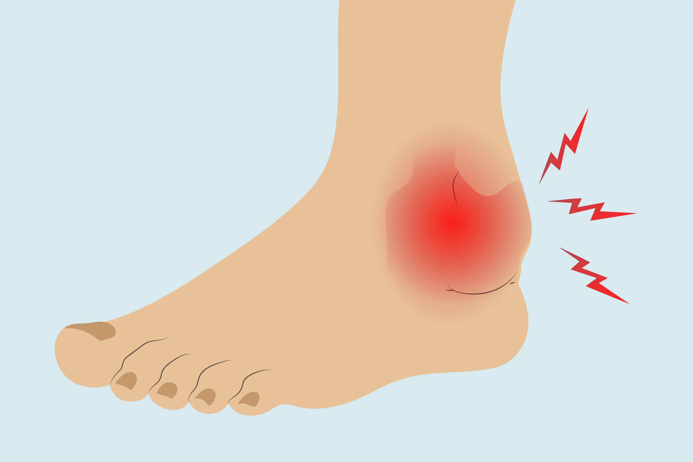
Arthritis in the Ankle: Treatments, Exercises, and Home Remedies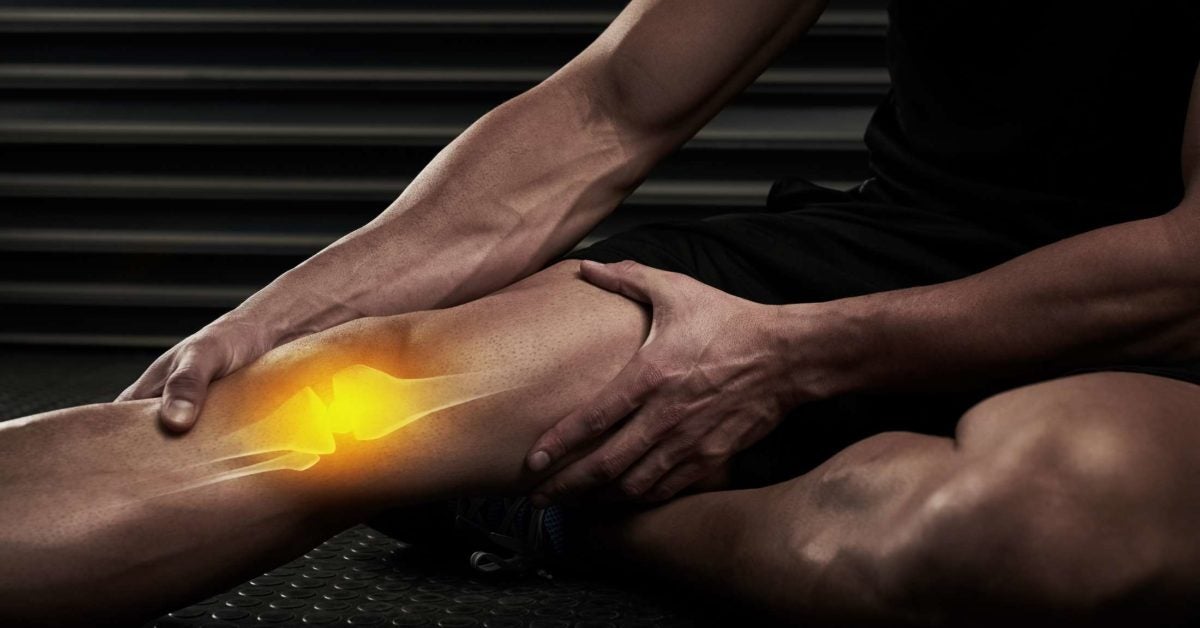
Inner knee pain: Treatment, exercises, and causes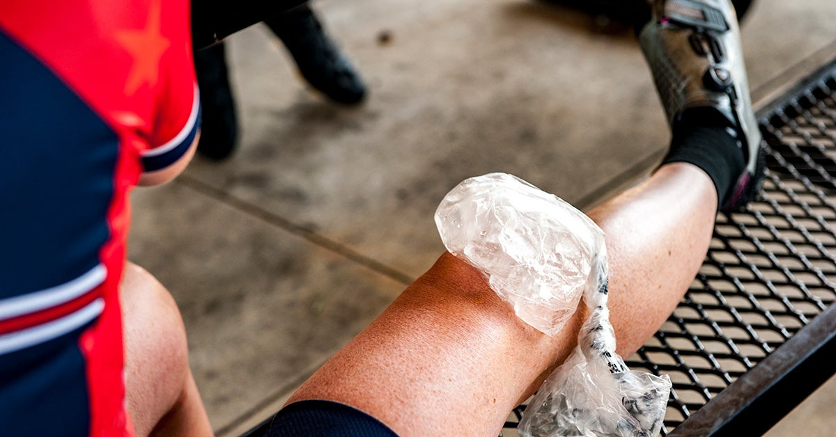
Sprained Knee Causes, Symptoms, Diagnosis, and Treatments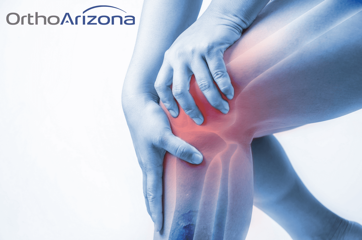
Should I Be Worried That My Joints Crack and Pop? - OrthoArizona | Complete Musculoskeletal Care/physical-therapy-after-ankle-fracture-2696531_v1-bf75f6bc0334467ab63054ce6f6845d2.gif)
Physical Therapy for an Ankle Fracture
What's causing pain on the inside of my knee?
Does ankle impingement require surgery? Improving range of motion without surgery – Caring Medical Florida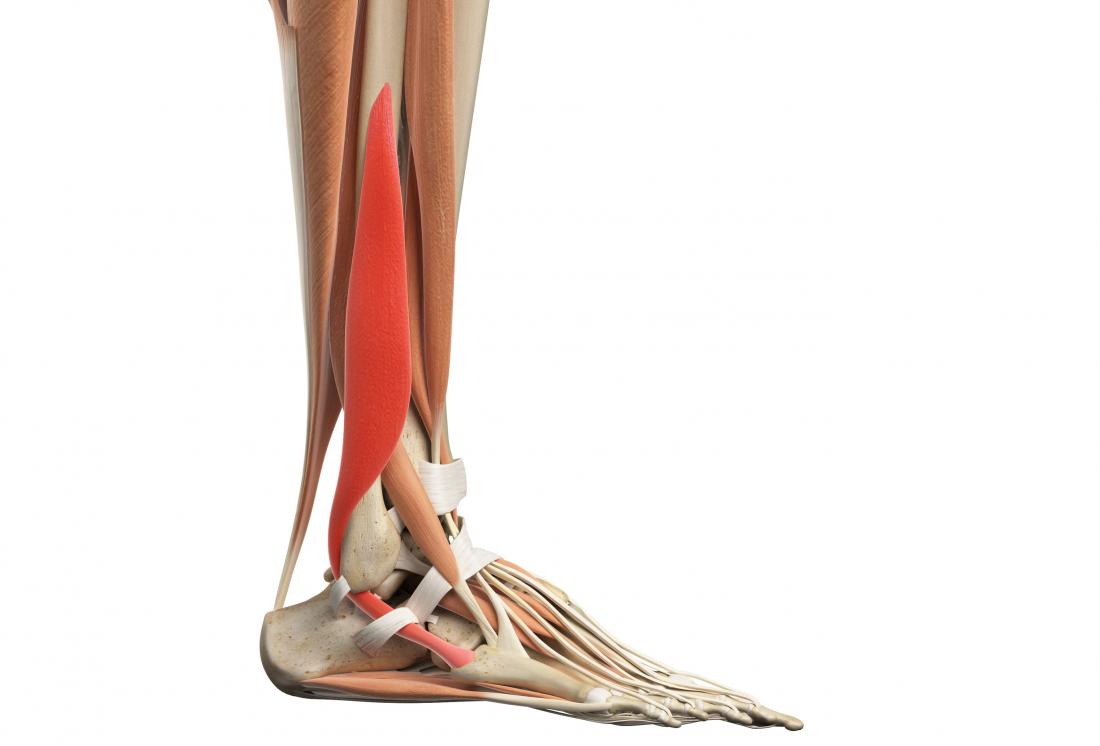
Plantar flexion: Function, anatomy, and injuries/kneetoankle1500-567837ce3df78ccc153421f5.jpg)
How to Do Knee to Ankle Pose (Agnistambhasana)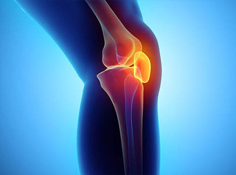
4 Ways to Fix Anterior Knee Pain from Cycling | ACTIVE
Tibia - Wikipedia
Ankle Sprain (Medial) | Upswing Health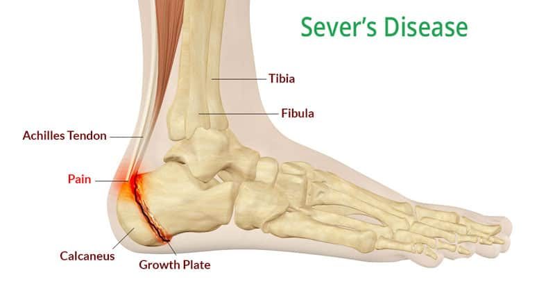
Sever's Disease and Osgood-Schlatter Disease - Back in Action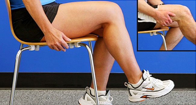
Exercises for Knee Osteoarthritis and Joint Pain
 the Knee Bone's Connected to the Ankle Bone - Regenexx Blog


/kneepainwhenrunning-58d2ae2b3df78c5162051537.jpg)


:background_color(FFFFFF):format(jpeg)/images/library/11077/175_176_177_178_179_tibia_extensors_nn.png)


/physical-therapy-after-ankle-fracture-2696531_v1-bf75f6bc0334467ab63054ce6f6845d2.gif)


/kneetoankle1500-567837ce3df78ccc153421f5.jpg)


Posting Komentar untuk "the knee is what to the ankle"